PLEASE PRINT OUT THESE PAGES FIRST AND THEN KEEP THE PRINTED TEXT BESIDE
YOU AS A GUIDE WHEN YOU LOAD AND RUN "NEURON". THIS WILL SAVE YOU HAVING
TO JUGGLE BETWEEN NEURON AND NETSCAPE WINDOWS ONCE YOU HAVE THE SIMULATION
LOADED.
To begin working with this chapter you should have downloaded and installed
Neuron, as described in Chapter 1.
-
This simulation brings together all of the concepts presented in Exercise
1 through 5. An unmyelinated axon, length 10,000 mm
and diameter 20 mm is simulated. A current electrode
is placed at one end of the axon (at position 0 mm)
and three voltage electrodes are placed at 0, 0.5 and 1.0 of the
length of the axon (at positions 0 mm, 5,000
mm and 10,000 mm).

-
Load the simulation, click on "IClamp" in the
bottom, center Window to bring up the parameters for the injected stimulating
current and then click on "Init & Run" in the bottom right-hand box
- you should see the action potential role across the left-hand graph.
The various graphs and windows that you will see are described below.
Hypotheses to be tested and related observations to be made:
-
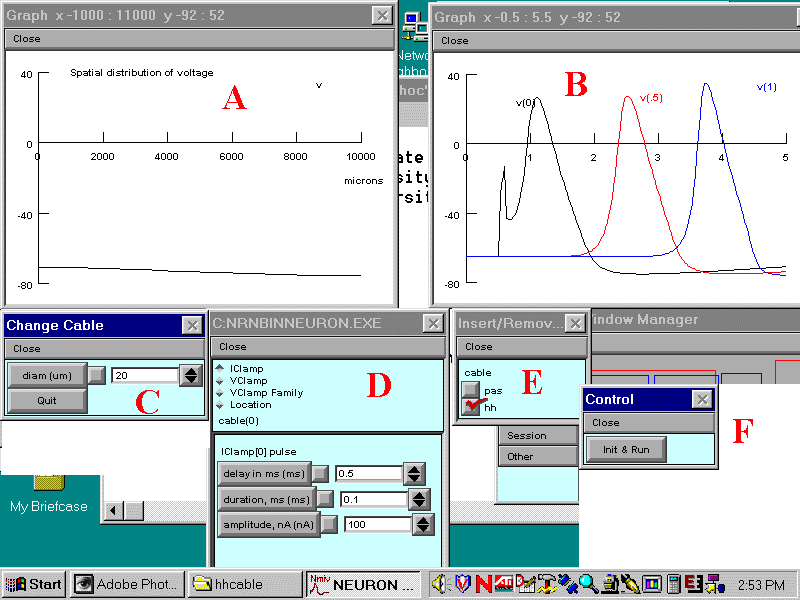
When you run the simulation observe and make sense of the correlation
between the movement of the action potential along the axon in Window A
and the voltage changes in Window B observed at the various fixed points
at which the electrodes are placed.
-
Distinguish between the depolarization created by the stimulating current
and the subsequent action potential. The effect of the stimulating current
occurs immediately at 0.5 ms and is only recorded at the 0 mm
voltage electrode.
-
At what velocity does the action potential travel (in mm/ms)?
Explain your reasoning.
-
What is the effect of removing the hh channels and replacing them
with voltage-independent (pas) channels. How far down the axon does the
depolarization produced by the stimulating current spread if there are
no voltage-dependent channels?
-
Alter the amplitude of the stimulating current - find the threshold for
triggering a traveling action potential.
-
What is the effect on action potential velocity of increasing the axon
diameter to 80 mm? (Hint: you will
also have to dramatically increase the amplitude of the injected current
to trigger an action potential in the larger axon - why?). Explain
the effect of diameter on action potential velocity given the discussion
in Exercise 4 of passive propagation.
When you have loaded the simulation you should see Windows arranged
as below. Click on the following letters: A B
C D E F
for a description of the function of each Window.
Return to Instructions

A. Graph of the calculated membrane potential (mV) as a function
of the distance (mm)
along the axon.
Return to letters
B. Graphs of membrane potential (mV) vs time (ms) recorded by
voltage electrodes placed at the various positions along the axon or dendrite.
Return to letters
C. The parameters of the axon:
diam - the axon or dendrite diameter
Return to letters
D. Stimulation Parameter Window: As shown, this allows
you to view and alter the amplitude, duration and delay of the stimulating
current injected into the current electrode.
You do not need to bother about the other buttons, which are not relevant
to this simulation.
Return to letters
E. This box determines the set of channels in the axon. Either a full
set of channels containing voltage-dependent and leakage channels can be
chosen by clicking on "hh" or a set of purely "passive" (i.e. voltage
independent) channels can be chosen by clicking on "pas".
F. The Run/Control Box - click on Init & Run to run a simulation.
Return to letters


